Figure 1.
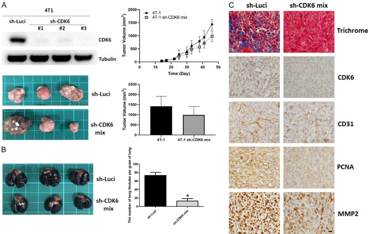
Knockdown of CDK6 reduces lung metastasis and malignant phenotype in vivo. A. Left panel, levels of CDK6 in parental and sh-CDK6 transfected 4T1 cells, and representative images of breast cancer tissue with high or low expression of CDK6 in BALB/c mice. Right panel, quantitative analysis of tumor growth and size 45 days after tumor cell injection. Comparisons utilized the 2-tailed Student t test (*P < 0.05). B. Lungs were perfused with India ink after the mice were euthanized. Left panel, representative images of lung nodules. Right panel, numbers of visualized nodules per gram of lung tissue compared using the 2-tailed Student t-test (*P < 0.05). C. Representative immunohistochemical staining of CDK6, PCNA, CD31 and MMP-2, and collagen (Masson’s trichrome stain) in tumors of parental and CDK6-deficient breast cancer tissues. Original magnification: 40 ×, scale bar: 20 μm.

