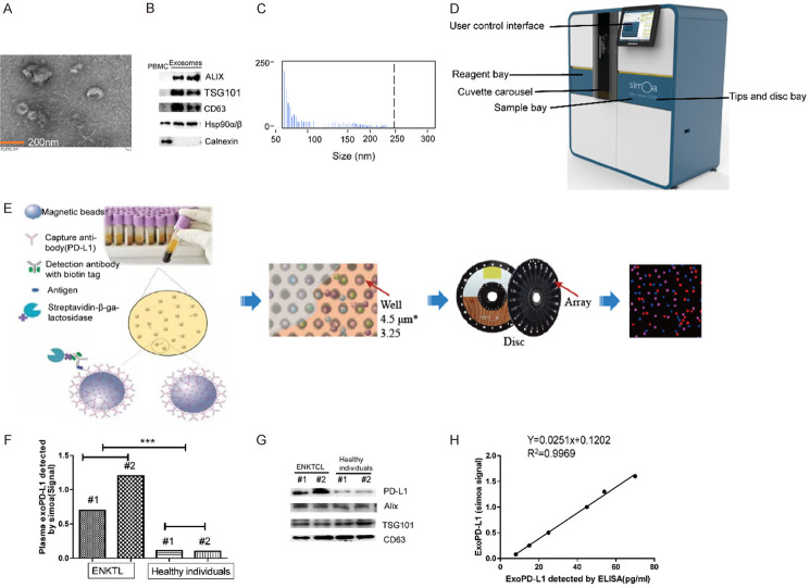
Figure 2.
(A-C) Characterization of isolated exosomes by transmission electron microscopy (TEM) (A), western blots (B) and size distribution (C). (D, E) Exosomes were captured on microscopic beads coated with anti-PD-L1 mAb and were then stained with labeled biotinylated anti-CD63 mAb, which could bind streptavidin-β-galactosidase (SBG) and catalyze substrate and produce signal after loading on the simoa disc array. (F, G) The plasma exoPD-L1 level of ENKTL patients and healthy individuals analyzed by western blots and simoa. (H) Linear relationship between the simoa signal and the plasma exoPD-L1 concentration detected by ELISA.

