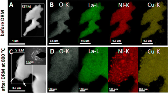
Figure 3.

Electron microscopy analysis of La2Ni0.9Cu0.1O4 in the initial and spent state after one DRM cycle (CO2:CH4:He = 1:1:3) in a total gas flow of 100 mL min–1 up to 800 °C for a total time of 2 h. Panels A and C: HAADF images, Panels B and D: EDX analysis of the O–K, La-L, Cu–K, and Ni–K intensities. Some exsolved Ni particles are marked by arrows in Panel C.
