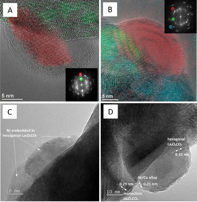
Figure 4.

Aberration-corrected high-resolution electron microscopy images of La2Ni0.9Cu0.1O4 (Panel A and C) and La1.8Ba0.2Ni0.9Cu0.1O4 (Panel B and D) in the spent state after one DRM cycle (CO2:CH4:He = 1:1:3) in a total gas flow of 100 mL min–1 at 800 °C for 2 h. Representative lattice fringes of relevant individual structures and phases have been color-coded. Color codes: Panel A – red: NiCu (200), green: hexagonal La2O2CO3 (002); Panel B – red: NiCu (111), green: hexagonal La2O2CO3 (202), blue: hexagonal La2O3 (011). The respective FFT patterns used for color-coding are shown as insets. The lattice fringes in Panels C and D correspond to hexagonal La2O2CO3 (102), NiCu (111), and monoclinic La2O2CO3 (023).
