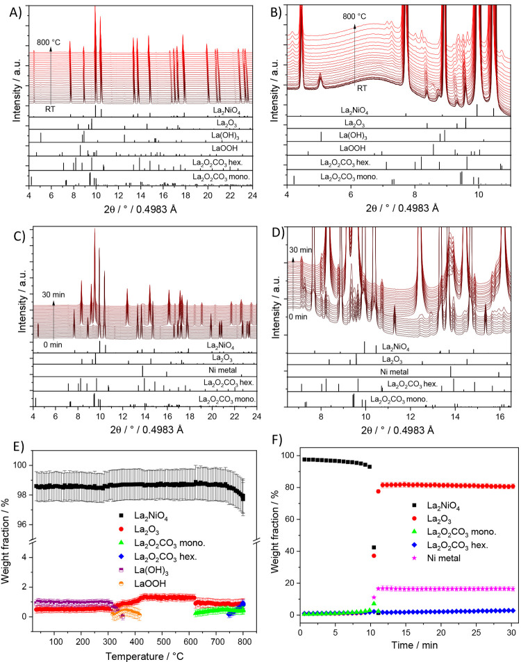
Figure 6.
Panel A: In situ collected XRD patterns of La2Ni0.8Cu0.2O4 during heating up to 800 °C under DRM conditions. Panel C: In situ collected XRD patterns of La2Ni0.8Cu0.2O4 during holding at 800 °C for 30 min under DRM conditions. Panels B and D focus on a narrower 2θ windows for closer analysis. The lower panels indicate the phase assignment to the respective reference structures. Panel E and F: Weight fractions of different crystalline phases formed during DRM as a function of temperature (E) and time at 800 °C (F) obtained by Rietveld refinement of the in situ collected XRD patterns of La2Ni0.8Cu0.2O4.

