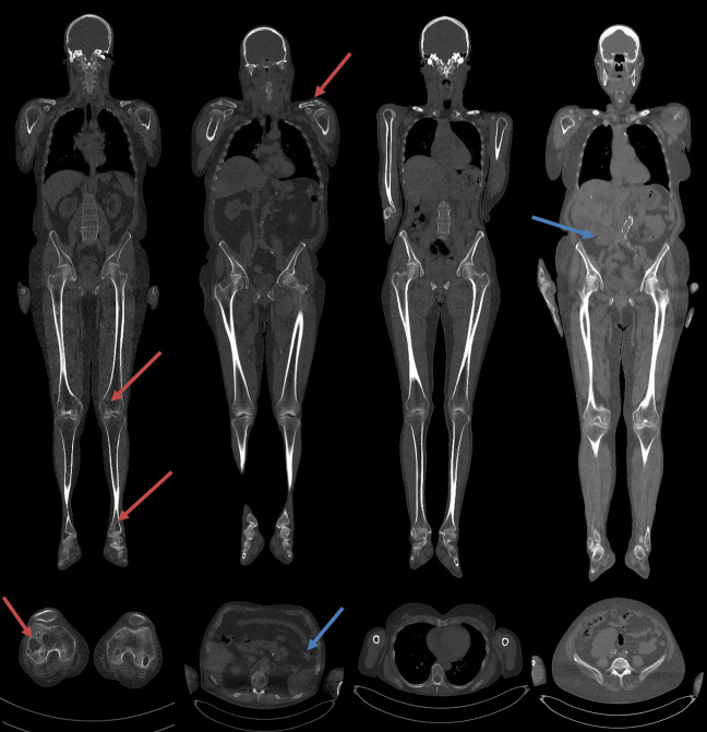
Figure 1.
Coronal and sagittal reconstructions from WBLDCT scan. From left to right, on top of the coronal MPR images and bottom the axial images, a patient showed the presence of osteolytic lesions, a patient also with extra-osseous findings (diverticulum), the patient showed no osteolytic lesions and patients only with extra-osseous findings (ectopic kidney). Blue arrows show the osteolytic lesion, the reds show the extra-osseous findings.

