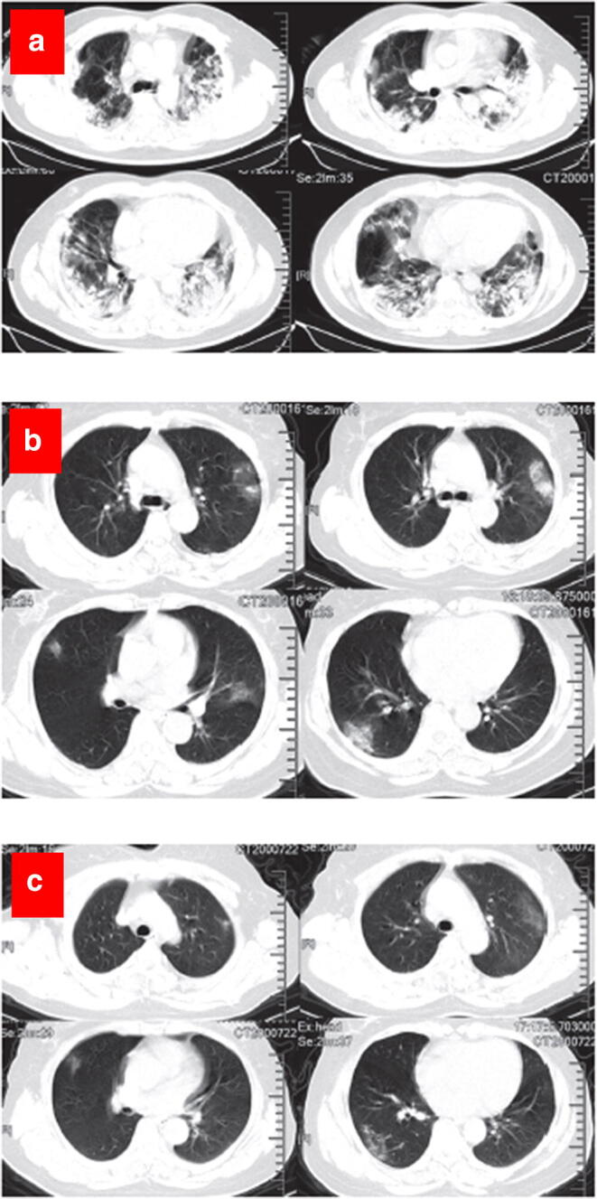
Fig. 9.

Representative chest CT images. a Transverse chest CT images from a 40-year-old man showing bilateral multiple lobular and subsegmental areas of consolidation on day 15 after symptom onset. b Transverse chest CT images from a 53-year-old woman showing bilateral ground-glass opacity and subsegmental areas of consolidation on day 8 after symptom onset. c Transverse chest CT images showing bilateral ground-glass opacity on day 12 after symptom onset. Reproduced with permission from (183)
