
Figure 1.
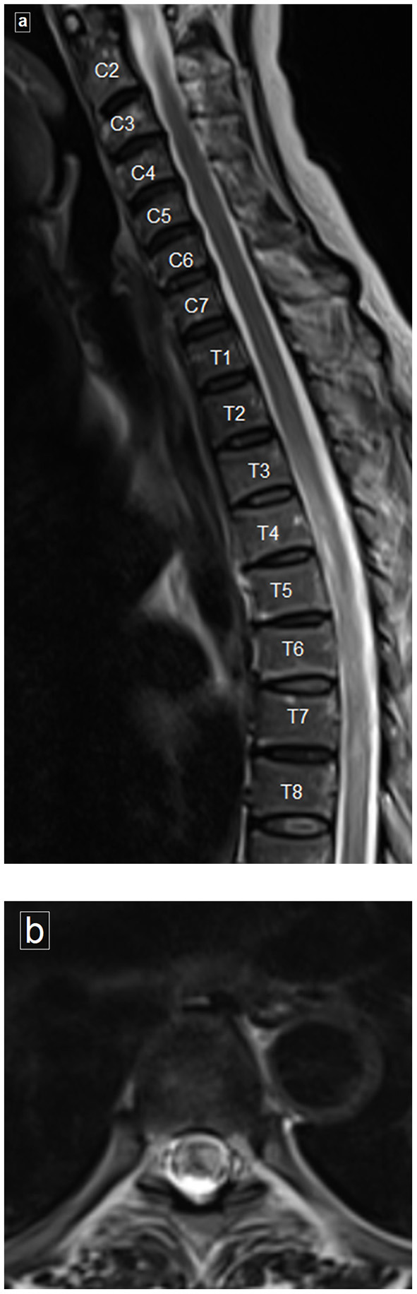
Magnetic resonance imaging (MRI) of spine. (a) Sagittal T2-weighted image of the cervical and dorsal spine reveals poorly delineated long segment of hyperintense signal, clearly evident from T4 to T8. (b) Axial T2-weighted image showing the hyperintense signal involving more than two thirds of the cross-sectional area of the cord.