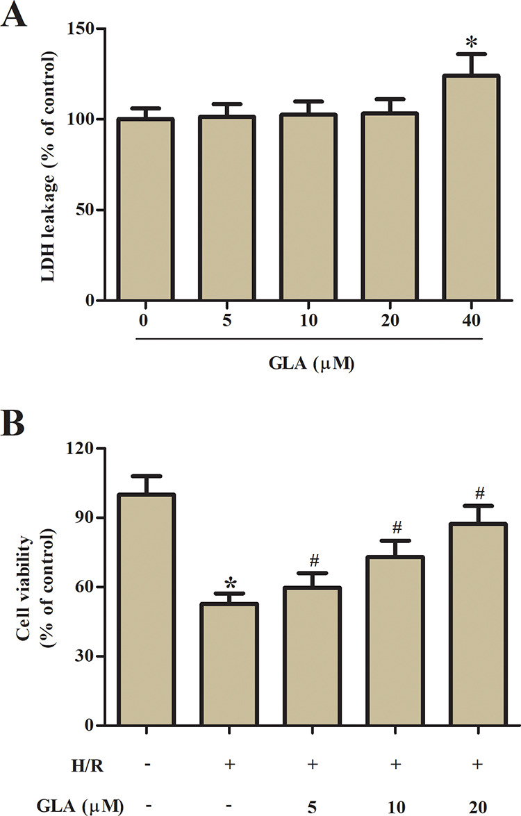
Fig. 1.

Effect of GLA on cell viability of H9c2 cells. (A) LDH leakage assay was performed to assess the cytotoxic effect of GLA on H9c2 cells after incubation with a series concentration of GLA (0, 5, 10, 20, and 40 μM) for 24 h. (B) MTT assay was performed to assess the protective effect of GLA on H/R-stimulated H9c2 cells. H9c2 cells were preincubated with GLA (5, 10, and 20 μM) for 2 h, and then subjected to H/R stimulation. *P < 0.05 indicates the significant difference compared with control H9c2 cells. # P < 0.05 indicates the significant difference compared with H/R-stimulated H9c2 cells. GLA: glaucocalyxin A; H/R: hypoxia/reoxygenation; LDH: lactate dehydrogenase; MTT: 3-(4,5-Dimethylthiazol-2-yl)-2,5-diphenyltetrazolium bromide.
