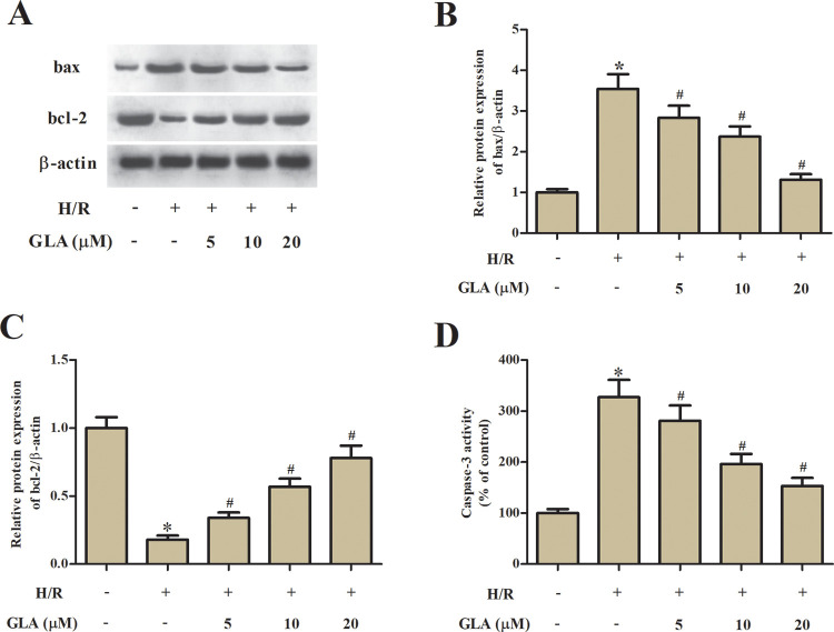
Fig. 3.
Effect of GLA on apoptosis in H9c2 cells. H9c2 cells were preincubated with GLA (5, 10, and 20 μM) for 2 h, and then subjected to H/R stimulation. (A) Western blot analysis was performed to detect the expression levels of apoptosis-related proteins, including bax and bcl-2. (B, C) Quantification analysis of bax and bcl-2. (D) Colorimetric method was used to determine the caspase-3 activity with the substrate peptide Ac-DEVD-pNA. *P < 0.05 indicates the significant difference compared with control H9c2 cells. # P < 0.05 indicates the significant difference compared with H/R-stimulated H9c2 cells. GLA: glaucocalyxin A; H/R: hypoxia/reoxygenation.

