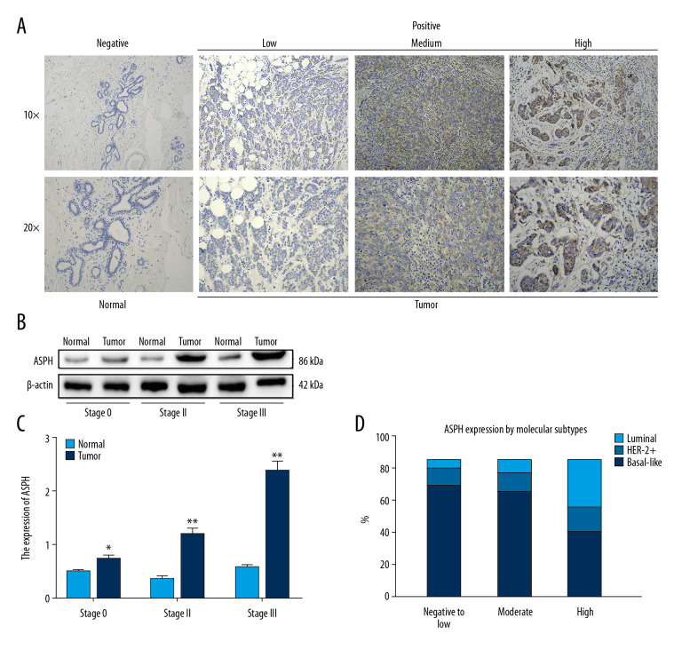
Figure 1.
Aspartate β-hydroxylase (ASPH) expression in breast cancer (BC). (A) Representative immunostaining for ASPH expression in negative normal breast tissue and in BC tissue showing weakly positive, moderately positive, and strongly positive staining. (B) Western blotting results of ASPH expression in tissues from patients with different stages of BC. (C) Quantitative analysis of ASPH expression in different stages of BC; n=30. (D) ASPH expression by molecular subtypes in BC. * P<0.05, ** P<0.01.

