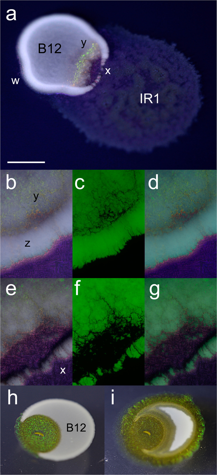
Fig. 2. IR1 invades and predates adjacent colonies of B12.

a Inoculation of IR1 adjacent to B12(pGFP) on ASWBFLow plates (ASWBLow agar supplemented with 0.5% w/v fucoidan), showing the result 10 h after contact between the spreading colony of IR1 and the static mass of B12. IR1 surrounds the B12 colony (w) and creates breaches (x) in the thicker edge of the B12 colony and a shift from dull purple/red SC typical of growth on ASWBFLow to green (y). IR1, IR1 colony; B12, B12 colony. b–d Images 4 h after contact with invading IR1. b Illumination from side showing white B12, with a thicker colony at the periphery (z) and SC from IR1 (bright pinpoints of colour including deep within the B12 colony) (y). c Fluorescence image showing GFP expressed by B12. d Merged (b) and (c). e–g are similar to b–d but after 9 h showing more extensive clearing of B12 cells and major breaches at periphery of the B12 colony (x). h and i show an experiment where B12 is inoculated in a droplet on to starvation medium, allowed to dry and then IR1 inoculated inside B12. h Result after 4 days showing expansion of the IR1 colony (IR1, showing predominantly green SC) to breach the periphery of the B12 colony (opaque white) from within. i Result of the same colony as (h) after 8 days showing progressive destruction of the B12 colony and movement around the periphery of B12 to engulf it. Scale bar indicates 0.4 mm for (a), 0.15 mm for (b–g) and 0.5 mm for (h) and (i).
