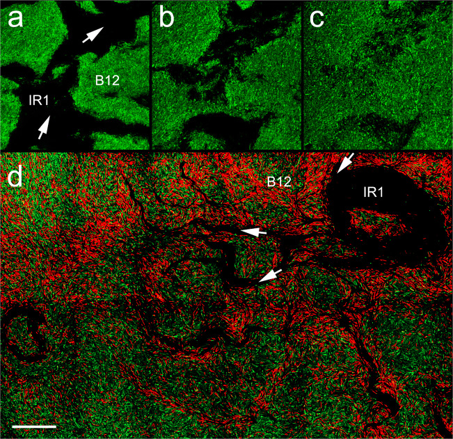
Fig. 3. Invasion of B12 by IR1 imaged by confocal microscopy.
a–c Three images taken from a Z-slice of a colony of B12(pGFP) during predation by IR1 (unstained, lines of advance shown with white arrows). From left to right the three slices show B12 cells at the agar surface, then 5, and 10 µm heights. d Overview image assembled from multiple contiguous images showing IR1 penetrating a colony of B12(pGFP). IR1 (not stained, visible as dark root-like regions but with an overall invasion route of top right to bottom left) is moving into a colony of GFP-expressing B12. White arrows show the direction of movement of some of the IR1 masses. Propidium iodide (red) is staining damaged cells (predominantly B12) within 20 μm of the major lines of advance of IR1. The scale bar in (d) indicates 50 µm when applied to (a–c) and 80 µm when applied to (d).

