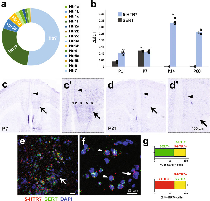
Fig. 1. 5-HTR7 expression in the mouse mPFC is developmentally regulated during the early postnatal period.
a Relative proportion of 5-HT receptor genes (Htr) expressed in PFC-SERT + neurons at P7, as revealed by RNA sequencing profiling of FACS-isolated SERTCre::EGFP neurons from the mPFC. b Time course of 5-HTR7 and SERT mRNA expression by q-PCR on mPFC tissue dissected at P1, P7, P14 and P60 (n = 3–4 per age). *p < 10−6 for SERT at P7 vs. all ages (F3,7 = 314.103; p < 10−7), and 5-HTR7 at P14 vs. P1–P7 (F3,7 = 129.497; p < 10−5) after ANOVA followed by Tukey’s test. c, d In situ hybridization of 5-HTR7 in the mPFC at P7 (c,c’) and P21 (d,d’) illustrating a laminar shift in the distribution of the receptor during the early postnatal period. Abundant expression of 5-HTR7 is present in layers 5–6 at early ages (arrows), however by P21 this expression remains only in superficial 2–3 layers (arrowheads). e, f RNAscope double fluorescent in situ hybridization in the mPFC at P7 (e, arrow), showing nuclear (DAPI, blue) co-localization of 5-HTR7 mRNA (red) with SERT mRNA (green) expression (arrowheads). Some of these mPFC neurons present 5-HTR7 mRNA but not SERT expression (f, arrow). g Quantitative analysis of the percentage of mPFC neurons co-expressing SERT (green) and 5-HTR7 (red) mRNAs.

