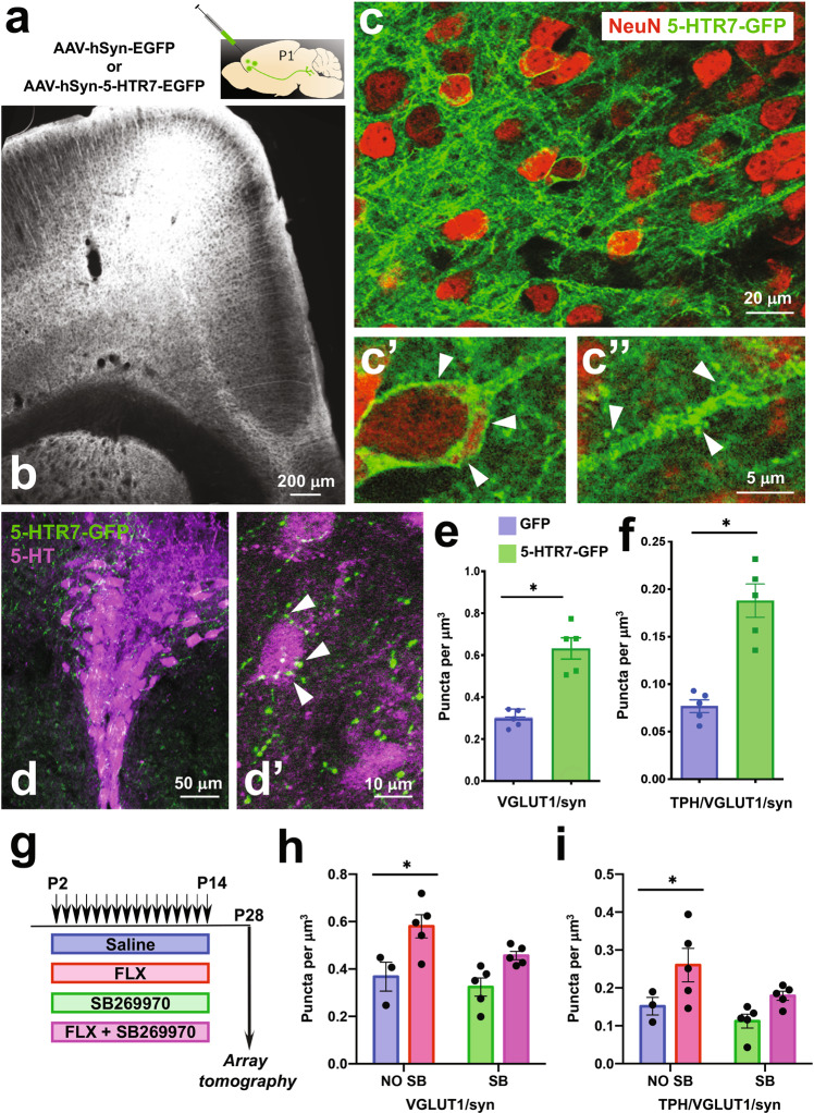
Fig. 3. Developmental role of 5-HTR7 in the synaptic wiring of the PFC-to-DRN circuit.
a, b Bilateral injections of the AAV-hSyn-5-HTR7-EGFP or AAV-hSyn-EGFP were carried out in the PFC of C57BL/6 mice at P1. Coronal section through the PFC at P12 shows heavy 5-HTR7-GFP expression in deep cortical layers (b). c High power confocal images of pyramidal neurons expressing the construct encoding 5-HTR7-GFP (green) with NeuN counterstaining (red). The 5-HTR7-GFP localizes at the cell membrane of pyramidal cell somas (c′) and dendritic processes including spines (c′′). 5-HTR7-GFP labeling is visible in axon terminals in the DRN (green) (d), often associated with TPH + neurons (5-HT, magenta) (d′). Quantification of VGLUT1/synapsin+ axonal boutons in the DRN of P28 mice that received injections of either sham (GFP; n = 5) or 5-HTR7-EGFP (n = 5) viral constructs in the PFC at P1. All VGLUT1/synapsin+ puncta (e), and VGLUT1/synapsin+ puncta in contact with TPH+ neurons (f) are shown. *p < 0.001. g–i Four groups of C57BL/6 mice were administered subcutaneously with saline (n = 3), FLX (10 mg/kg/day; n = 5), FLX (10 mg/kg/day) + SB269970 (10 mg/kg/12 h) (n = 5) or SB269970 (10 mg/kg/12 h; n = 5) during the critical period (P2–P14) . Array tomography quantitative analyses were carried out in the DRN at P28 in all four experimental groups (g). Density of VGLUT1/synapsin+ puncta (h) [total number of puncta analyzed for saline (21,510), FLX (45,166), FLX+ SB269970 (35,215) and SB269970 alone (26,892)], and VGLUT1/synapsin+ puncta related to TPH + neurons (i) [total number of puncta analyzed for saline (7059), FLX (20,011), FLX+ SB269970 (14,563) and SB269970 alone (8907)]. *p < 0.001 and p < 0.01 for h and i, respectively, FLX vs. all treatments.

