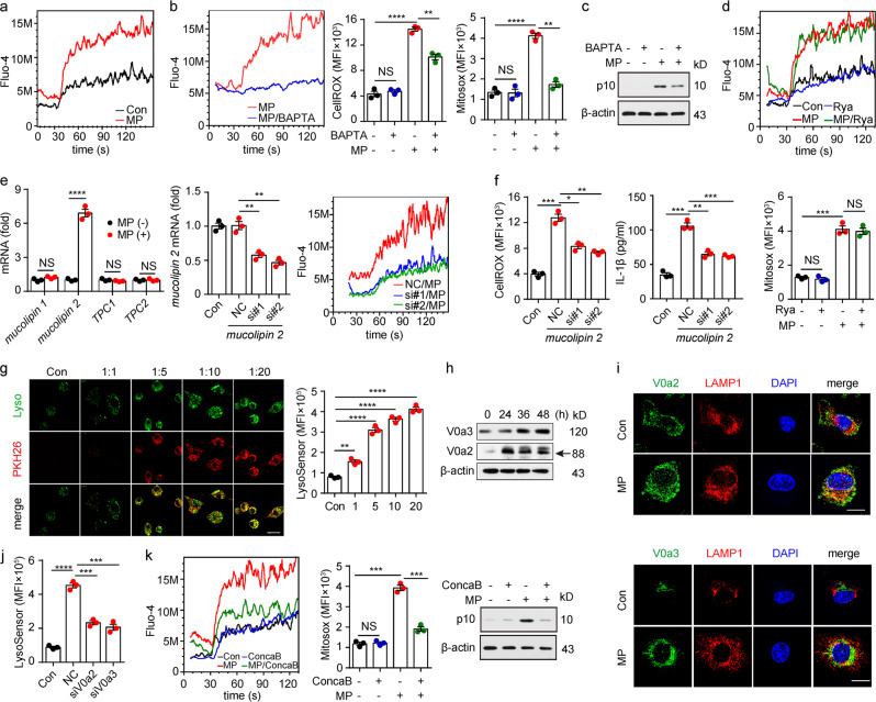
Fig. 5.
Lysosomal calcium release by L-MPs causes mitochondrial ROS production. a Mouse BMDMs treated with or without Lewis-MPs were loaded with the fluorescent Ca2+ indicator Fluo-4/AM. The Fluo-4 fluorescence intensity of the macrophages was measured via flow cytometry. Cells were stimulated with ionomycin (100 μM, 30 s). b, c Mouse BMDMs were treated with Lewis-MPs with or without BAPTA for 24 h, and then, the Ca2+ (b, left), ROS (b, middle), mitochondrial ROS (b, right) and active caspase-1 (c) levels of the macrophages were measured with flow cytometry or western blots. d Mouse BMDMs were treated with Lewis-MPs with or without ryanodine (Rya). After 24 h, the Ca2+ (upper) and mitochondrial ROS (bottom) levels of macrophages were measured via flow cytometry. e, f Mouse BMDMs were treated with or without Lewis-MPs. After 2 h, the cells were collected, and RNA was extracted for mRNA analysis of mucolipin 1, mucolipin 2, TPC1 and TPC2 by real-time PCR (e, left). The BMDMs were transfected with mucolipin 2 siRNAs, and the silencing efficiency of the siRNAs was detected by real-time PCR (e, middle). The BMDMs transfected with mucolipin 2 siRNAs were treated with Lewis-MPs. After 24 h, the Ca2+ (e, right) and ROS (f, left) levels of the macrophages were measured via flow cytometry. After 72 h, the culture medium was collected, and IL-1β expression was analyzed by ELISAs (f, right). g Mouse BMDMs were incubated with PKH26-labeled Lewis-MPs at different doses. After 12 h, the BMDMs were stained with LysoSensor. Then, the cells were observed under a two-photon confocal microscope. Scale bar, 20 µm (left). The flt1 LysoSensor fluorescence intensity of the macrophages was measured via flow cytometry (right). h Mouse BMDMs were treated with Lewis-MPs at different times. Then, the expression of V0a2 and V0a3 was detected via western blots. i Mouse BMDMs were treated with Lewis-MPs for 24 h. Immunofluorescence of V0a2 (green, upper), V0a3 (green, bottom) and LAMP1 (red) in the control and Lewis-MPs groups was assessed with two-photon confocal microscopy. Scale bar, 20 µm. j Mouse BMDMs were transfected with V0a2 or V0a3 siRNAs and then treated with Lewis-MPs. After 12 h, the cells were stained with LysoSensor, and the flt1 LysoSensor fluorescence intensity of the macrophages was measured by flow cytometry. k Mouse BMDMs were treated with Lewis-MPs with or without concanamycin B (ConcaB). After 24 h, the cells were stained with Fluo-4 (left) or MitoSox (middle). Then, the Fluo-4 and MitoSox fluorescence intensity of the macrophages was measured by flow cytometry. The active caspase-1 levels were detected by western blots (right). Error bars indicate the mean ± SEM; n = 3 independent experiments. *P < 0.05, **P < 0.01, ***P < 0.001, ****P < 0.0001

