Fig. 1.
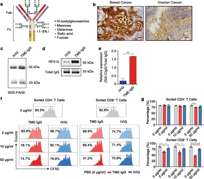
Purified TME-IgG containing abundant SIA-CIgG can directly inhibit T-cell proliferation. a The structure of the sugar moiety that is linked to the Fc fragment of intravenous immunoglobulin (IVIG) with enhanced anti-inflammatory activity. The structures of sialylated N-glycans located at the Asn162 site of SIA-CIgG Fab fragments and classic sialylated N-glycans attached to Asn297 in IgG Fc fragments are shown. Variable residues such as N-acetylglucosamine (blue square), mannose (green circle), galactose (yellow circle), sialic acid (purple diamonds), and fucose (red triangle) are shown. The classic sialylated N-glycans attached to Asn297 in IgG were present in ~1–5% of the total serum IgG pool. b Immunohistochemical staining for SIA-CIgG in the TME of human breast and ovarian cancer tissue samples using RP215. Scale bars, 100 μm. c Identification of purified TME-IgG using SDS-PAGE. IVIG was used as a control. d SIA-CIgG in TME-IgG detected by western blotting using RP215, and total IgG estimated by a commercial anti-IgG antibody. e Expression of SIA-CIgG relative to that of the total IgG evaluated as in (d) and calculated with the average gray value of three measurements for each band. f Proliferation of PBS-, TME-IgG-, and IVIG-treated CD4+ and CD8+ T cells. The doses of TME-IgG or IVIG were 0 μg/ml, 10 μg/ml, and 50 μg/ml. Cells were sorted by flow cytometry, labeled with carboxyfluorescein succinimidyl ester (CFSE), and then activated for 72 h using precoated 3 μg/ml anti-CD3 and 1 μg/ml anti-CD28 mAbs. g Proportion and statistical significance of proliferating CD4+ and CD8+ T cells sorted by flow cytometry and treated with TME-IgG or IVIG as in (f) (n = 3 each group). IVIG intravenous immunoglobulin, TME-IgG tumor microenvironment IgG. Small horizontal lines (e, g) indicate the mean ( ± s.d.). *P ≤ 0.05, **P ≤ 0.01, and ****P ≤ 0.0001 (two-tailed Student’s t test for unpaired data). Data are from one experiment that was representative of three (c–g) independent experiments with similar results. Also see Supplementary Fig. S1.

