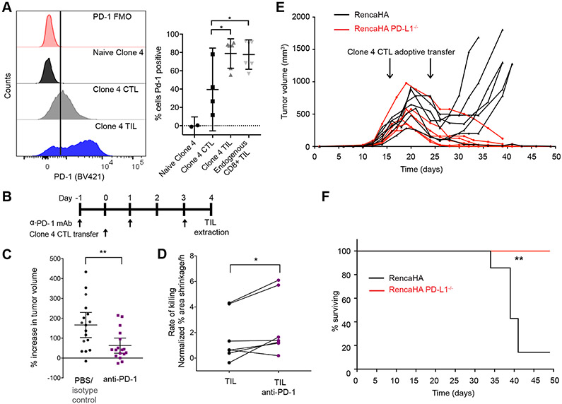
Fig. 7. Loss of PD-1 engagement in vivo improves TIL killing ability.
(A) Flow cytometric analysis of the cell surface expression of PD-1 on naïve and primed Clone 4 CTLs, Clone 4 TILs, and endogenous CD8+ TILs. Left: Histograms are representative of at least two independent experiments. Right: Data are means ± 95% confidence interval. (B and C) Analysis of subcutaneous RencaHA tumor growth in mice after treatment with antibody against PD-1 and adoptive transfer of Clone 4 CTLs, as indicated. The percentage increase in tumor volume within 5 days of the initiation of antibody treatment for 17 mice/condition with means ± 95% confidence interval are from five independent experiments. (D) Fluorescence microscopy analysis of the killing of Renca+HA cells in vitro by HA-specific TILs from untreated control mice or mice treated with antibodies against PD-1. Average Renca+HA cell death rate data with means ± 95% confidence interval are from six independent experiments. (E and F) Analysis of subcutaneous RencaHA and RencaHA PD-L1−/− tumor growth in mice after adoptive transfer of HA-specific CTLs at the indicated times. Tumor volume (E) and survival (F) data of 7 RencaHA and 6 RencaHA PD-L1−/− tumor-bearing mice are pooled from two independent experiments. *P < 0.05 and **P < 0.01 by one-way ANOVA (A), Mann Whitney u-test (C), paired Student’s t-test (D), or Mantel-Cox test (F).

