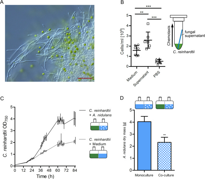
Fig. 5. Association of A. nidulans with C. reinhardtii.
a Microscopy picture showing C. reinhardtii cells (green) in mycelium of A. nidulans. Scale bar: 50 µm. b Numbers of C. reinhardtii cells found in capillaries filled with culture supernatant of A. nidulans, medium or PBS. ** represents P ≤ 0.01, ***P ≤ 0.001, calculated from at least three biological replicates; error bars indicate SDs. c Optical density of C. reinhardtii cells reached after co-cultivation with A. nidulans in co-cultivation chambers, separated by a PVDF membrane pore size 0.1 µm. d Dry masses of A. nidulans grown in mono- and co-culture.

