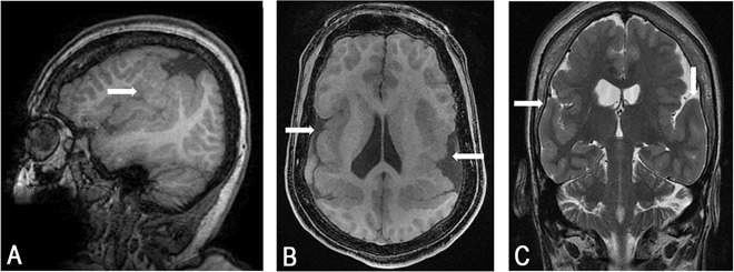
Fig. 1. Magnetic resonance imaging of the patient brain showing bilateral perisylvian polymicrogyria (arrows).
Representative sagittal fast spoiled gradient echo (FSPGR) image (a), axial FSPGR image (b) and coronal fast spin echo (FSE) T2 image (c) sections are shown. Notice asymmetry of location and extent of abnormal cortex on both hemispheres and asymmetric ventricles possibly explaining the patients’ hemiparesis in addition to her opercular symptoms and signs.

