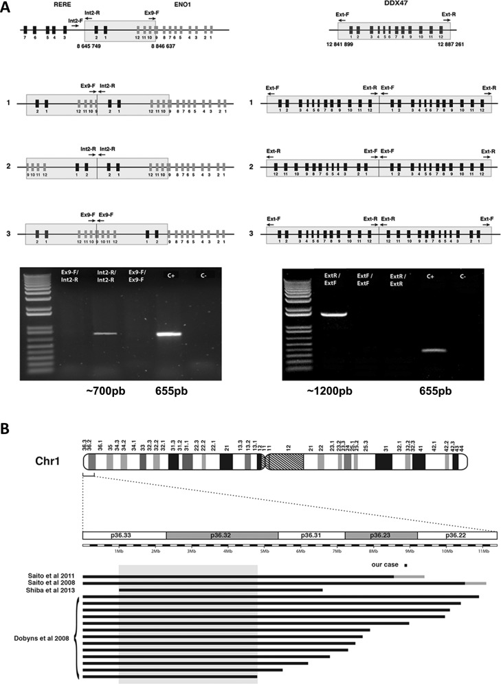
Fig. 3. Structure of the duplicated segments and comparison with published cases.
a Determination of the orientation of the duplicated segments. The three possible configurations are shown: in tandem (1), in mirror at the distal side (2) or in mirror at the proximal side (3). Arrows indicate orientation of PCR primers. Positive PCR amplifications reveal that the 1p36 duplication occurred in mirror while it occurred in tandem in 12p13.1. The positive control corresponds to the amplification of a portion of the TBC1D24 gene on chromosome 16, to check for proper PCR conditions. b Location of the 1p36 duplicated region in the reported patient with respect to previously reported and precisely characterized 1p36 deletion cases with PMG. Deleted regions are represented as black lines, gray lines identify uncertainly delimited deletions. The gray box denotes the previously published minimal critical regions for PGM, located between 1 and 4.8 Mb on chromosome 1.

