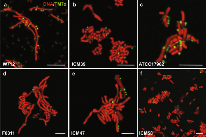
Fig. 4. TM7x localization on ICM58.
FISH imaging was carried out for all permissive (a–c, see Fig. S7) and all nonpermissive (d–f) bacterial hosts. TM7x (green) was visualized using a Saccharibacteria-specific DNA probe tagged with the Cy5 fluorescent molecule. The host bacteria were visualized by universal nucleic acid stain syto9, which also stains TM7x. Only sample strains are shown in this figure, and the complete set can be found in Fig. S7, including a few of the resistant strains visualized by FISH. Scale bars are 5 μm.

