FIGURE 4.
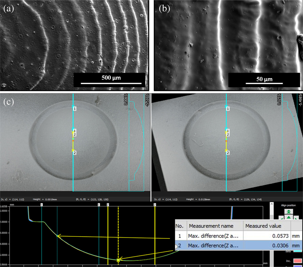
Characterization of corneal support scaffold (a, b) SEM of PDMS corneas derived from support scaffold made of Dental SG Resin showing higher anterior surface resolution of SLA printed scaffold. (c) Brightfield images were captured for two different wells on the same scaffold to confirm the radius of the well and variation between two wells

