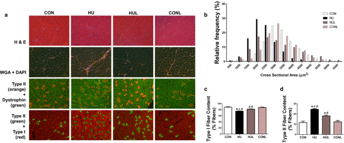
FIGURE 1.

AT1R blockade mitigates soleus muscle fiber remodeling due to one week of hindlimb unloading. A (top row), representative images of hematoxylin & eosin (H&E) stained soleus muscles; (second row), representative images of muscles stained for wheat germ agglutinin (WGA) and DAPI. (third row), representative immunofluorescence images of soleus muscles stained for type II muscle fibers (orange) and dystrophin (green); (bottom row), images of soleus muscles stained for type II muscle fibers (red) and type I muscle fibers (green). Scale bar = 100 µm. B, Distribution of muscle fibers according to fiber cross‐sectional area (CSA) for controls (CON), hindlimb unloaded (HU), hindlimb unloaded + Losartan (HUL), and control + Losartan (CONL) (n = 7/ group). C, percentage of soleus muscle fibers that are type I fibers. D, percentage of muscle fibers that are type II fibers. Values are means ± SEM. Letters indicate groups are significantly different from each other (p < .05): aindicates different from CON; cindicates different from HUL; dindicates different from CONL
