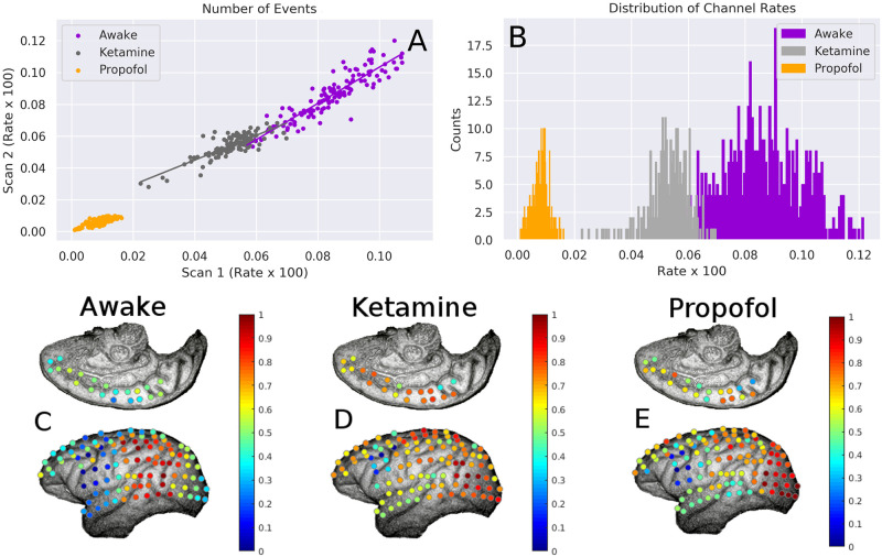
Fig 6. Changes in channel activity levels between conditions.
A: For each condition, the correlation between channel activity (firing rate × 100) between two scans. In all conditions, it is apparent that those channels that are more activity in one scan are similarly active during different scans within the same condition. This suggests that brain dynamics are consistent within conditions. B: histograms of the the channel rate × 100 for all three conditions. It is apparent that propofol decreases the overall channel firing rate more that ketamine, although both have a lower firing rate than the awake condition. C-E: normalized channel activity rates projected onto the recording array. It is clear that propofol and ketamine disrupt the activity-rate structure that exists in the awake condition. Original images taken from the NeuroTycho wiki: http://neurotycho.org/spatial-map-ecog-array-task.

