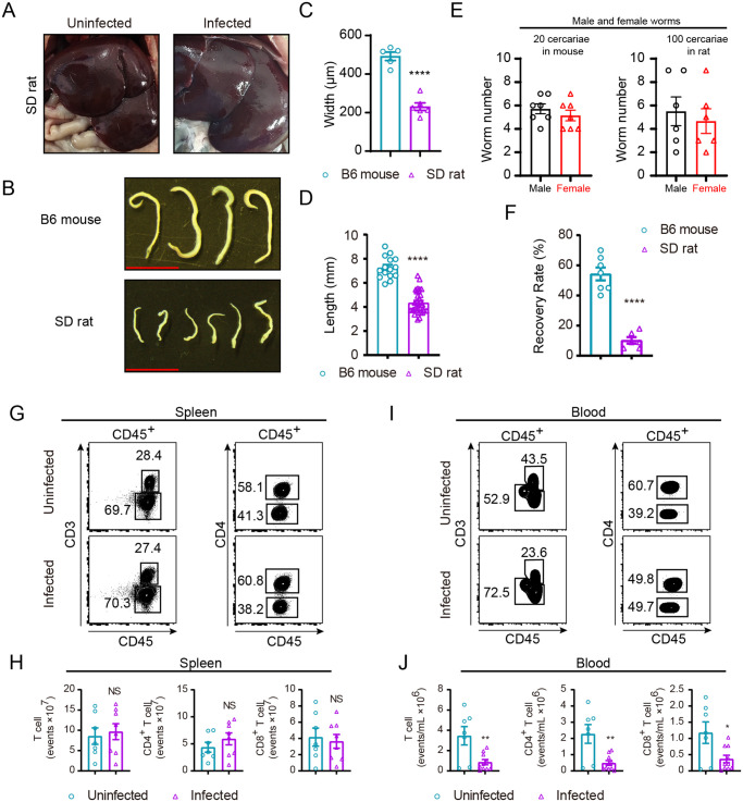
Fig 2. Immunopathologic characteristics of SD rat after S. japonicum infection.
(A) S. japonicum infection does not cause overt liver granuloma in SD rat, liver of uninfected SD rat (left) and liver of infected (right). (B) Representative micrographs of worms collected from portal and mesenteric veins of B6 mouse (left) and SD rat (right) both infected with S. japonicum for 6 weeks (Scale bar, 5 mm). (C) Width of worms from B6 mice and SD rats at week 6 post-challenge was measured based on digital micrographs. Data are from two experiments (B6 mouse, n = 5; SD rat, n = 6). (D) Length of worms harvested from portal and mesenteric veins of infected B6 mouse and SD rat was determined. Data are from two independent experiments (B6 mouse, n = 15; SD rat, n = 33). (E) Number of male and female worms recovered from B6 mouse and SD rat at 6-week post-challenge. (F) Percentage of worm recovery from portal and mesenteric veins of infected B6 mouse and SD rat, presented from two experiments (B6 mouse, n = 7; SD rat, n = 6). (G-I) Representative FACS plots of immune cells in spleen (G) or peripheral blood (I) of SD rat under uninfected and infected conditions. (H, J) Absolute number of total T cells, CD4+ or CD8+ T cells in G and I. Data are from two independent experiments (H, uninfected, n = 7; infected, n = 8. J, uninfected, n = 7; infected, n = 10). The following fluorochrome-tagged antibodies were used: anti-rat CD45 -eFluor450, anti-rat CD3-APC, anti-rat CD4-APC/Cyanine7, anti-rat CD8α-PE. Data represent the mean ± s.e.m. Statistical significance was assessed by unpaired Student’s t-test or non-parametric unpaired Mann-Whitney test and indicated by * P<0.05, ** P<0.01, **** P<0.0001, NS, non-significant.

