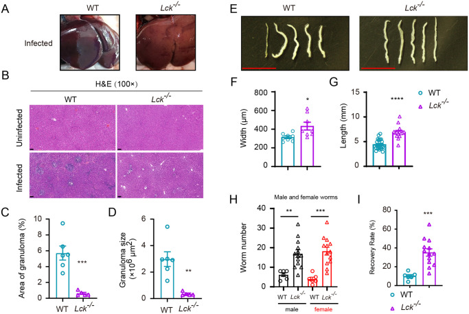
Fig 4. Immunopathologic analysis of Lck−/− SD rat following schistosome infection.
(A) S. japonicum skin infection does not cause noticeable liver granuloma in Lck−/− SD rat (right) compared to the liver of infected WT rat control (left). (B) H&E staining of liver sections from uninfected or infected WT SD rat and Lck−/− SD rat. (Original magnification, ×100; Scale bar, 100 μm). (C, D) Granuloma size was determined based on H&E staining of liver sections using CaseViewer software (WT rat, n = 6; Lck−/− rat, n = 5). (E) Representative micrographs of S. japonicum recovered from portal and mesenteric veins of infected WT SD rat (left) or Lck−/− SD rat (right) at 42 days after infection (Scale bar, 5 mm). (F) Width of worms from WT rat and Lck−/− rat at week 6 post-challenge was measured based on digital micrographs (worms from WT rats, n=8; worms from Lck−/− rat, n = 8). (D) Length of worms harvested from infected WT SD rat and Lck−/− SD rat. Data are from two independent experiments (worms from WT rat, n = 25; worms from Lck−/− rat, n = 15). (H) Number of male and female worms recovered from WT and Lck−/− SD rat at 6-week post-infection. (I) The recovery rate of S. japonicum collected from infected WT rat and Lck−/− SD rat. Data are from two independent experiments (WT rat, n = 6; Lck−/− SD rat, n = 13). Data represent the mean ± s.e.m. Statistical significance was assessed by unpaired Student’s t-test or non-parametric unpaired Mann-Whitney test and indicated by * P<0.05, ** P<0.01, *** P<0.001, **** P<0.0001.

