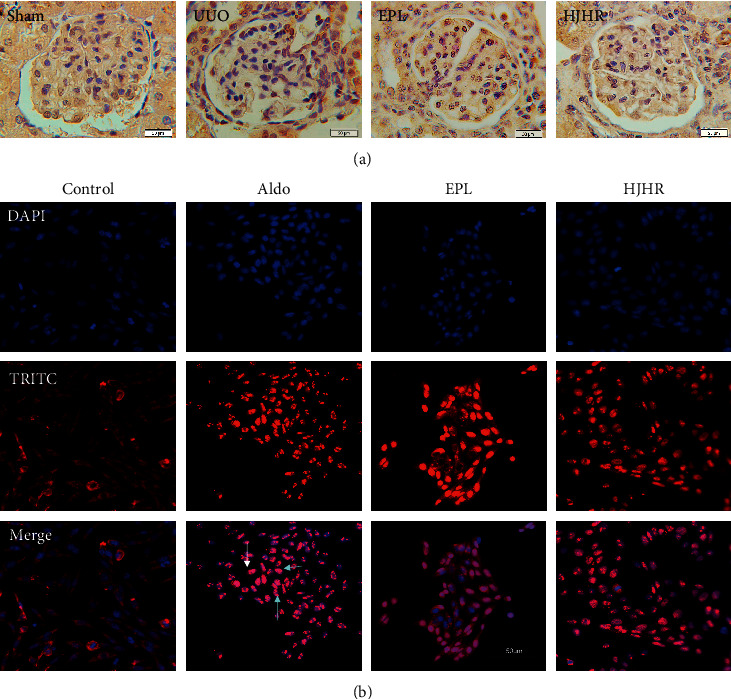
Figure 3.

(a) Expression of mineralocorticoid receptor (MR). Rat contralateral glomerular sections were analysed by immunohistochemistry using antibodies against MR. (b) Human mesangial cells were treated for 24 h with vehicle or 1 μM of aldosterone at 37°C. Cells were fixed and analysed by EVOS using antibodies against MR (red staining). Cells were also stained with DAPI (nucleus, blue staining).
