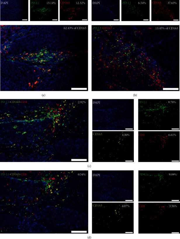
Figure 2.

CD4+ TILs colocalize with PD-L1+CD163+ TAMs. Representative multicolor immunofluorescence (multi-IF) images showing (a) high and (b) low colocalization of PD-L1+ cells with CD163+ TAMs. Scale bar, 100 μm. Representative multi-IF images showing costaining of (c) CD4+PD-L1+CD163+ TILs and (d) CD8+PD-L1+CD163+ TILs in serial sections of the same specimen. Cells were counterstained with DAPI (blue, nucleus). Scale bar, 100 μm.
