Figure 3.

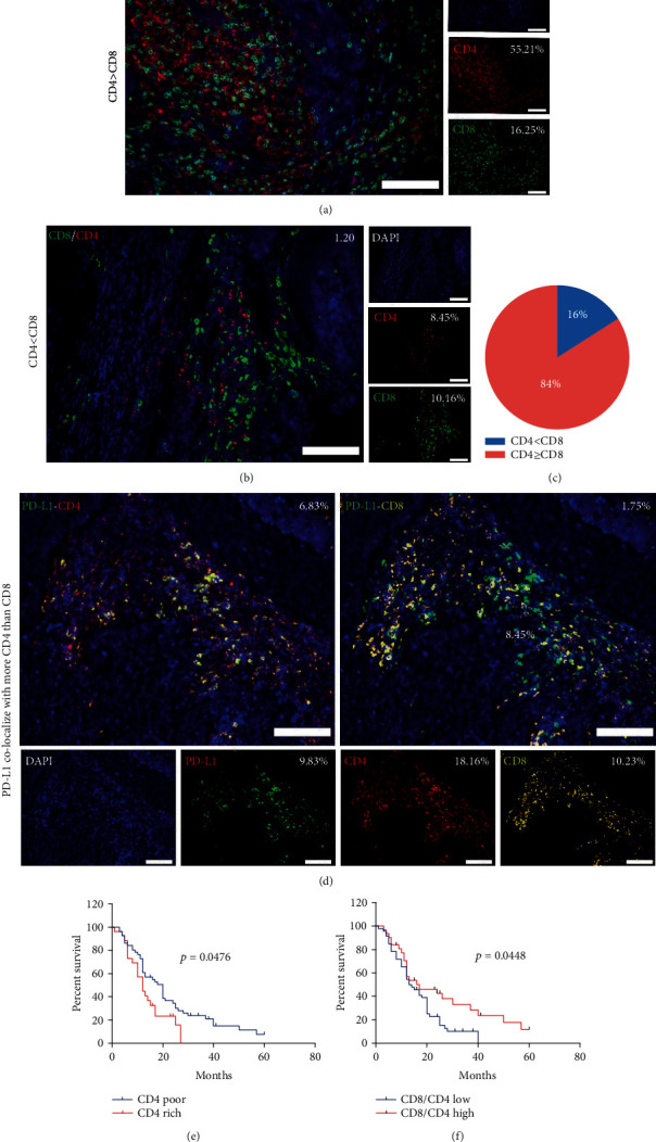
CD4+ TILs colocalize with PD-L1+ cells and contribute to poor survival. (a) Representative multi-IF images showing a sample with more CD4+ TILs than CD8+ TILs in the stroma. Scale bar, 100 μm. (b) Representative multi-IF images showing a sample with more CD8+ TILs than CD4+ TILs in the stroma. Scale bar, 100 μm. (c) A pie chart was plotted according to IHC staining results showing 84% of patients have more CD4+ TILs than CD8+ TILs in the stroma. (d) Representative multi-IF images showing a sample with PD-L1+ cells colocalizing more with CD4+ TILs than with CD8+ TILs in the stroma. Scale bar, 100 μm. (e) The Kaplan–Meier survival analysis showing patients with elevated levels of CD4+ TILs (red line, n = 26) have poor OS, compared to patients with low levels of CD4+ TILs (blue line, n = 51). (f) The Kaplan–Meier survival analysis showing patients with a low CD8/CD4 ratio (blue line, n = 46) have poor OS, compared to patients with a high CD8/CD4 ratio (red line, n = 31).
