
Fig. 4.
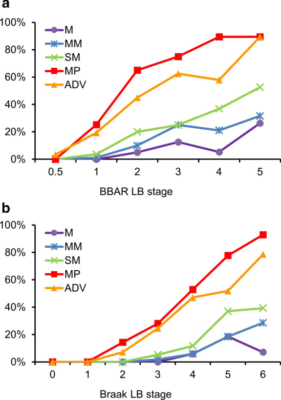
Lewy pathology in the esophageal wall at different BBAR and Braak LB stages. a BBAR LB stages; The positive rate of Lewy pathology increased with BBAR LB stage. The highest rates were found in the MP and ADV (mean rates of 41.6% and 33.1% respectively), reaching 89.5% at stage 5. Lewy pathology was observed less in the M and MM. b Braak LB stages; The positivity of Lewy pathology also increased with Braak LB stage. The highest rates were found in the MP, reaching 92.9% at stage 6. Lewy pathology was observed less in the M and MM. Details are summarized in Table 4a, b. BBAR the Brain Bank for Aging Research, LB Lewy body, M mucosa, MM muscularis mucosa, SM submucosa, MP muscularis propria, ADV adventitia