Figure 4.
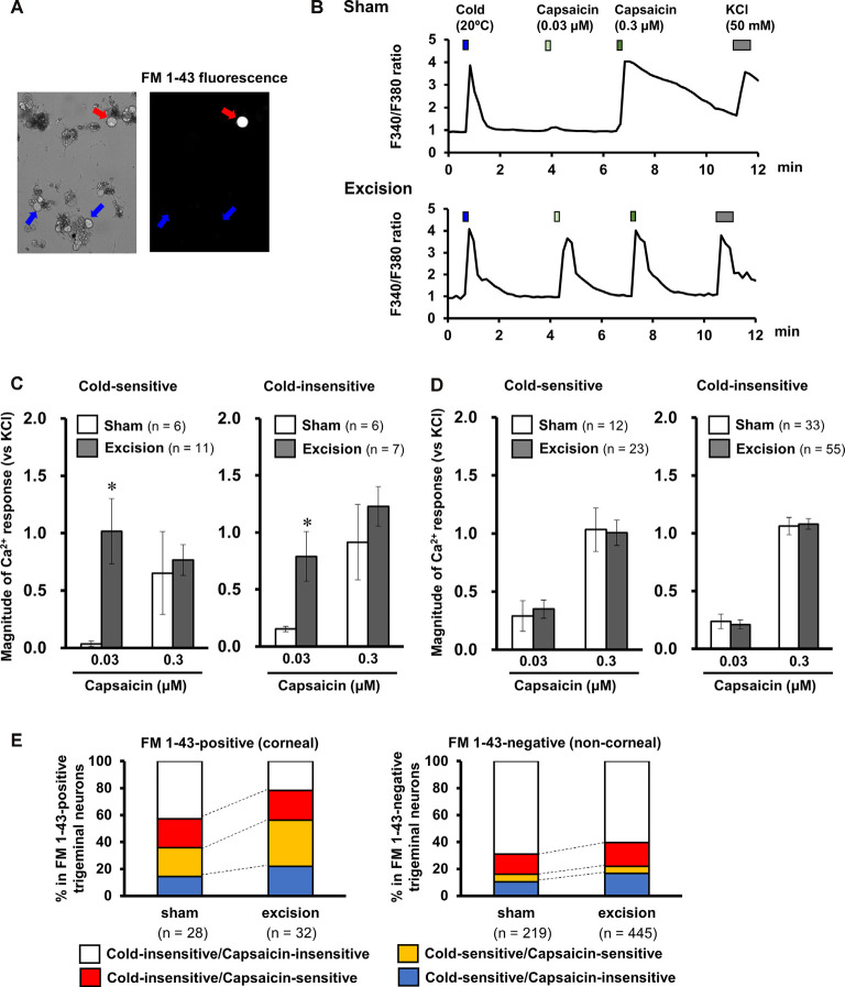
Effect of lacrimal gland excision on capsaicin-induced intracellular calcium response in the corneal and non-corneal trigeminal ganglion neurons. (A) Representative image of an FM 1-43-positive corneal neuron (red arrow) and FM 1-43 negative non-corneal neuron (blue arrows). (B) Representative trace of the Fura-2 fluorescence ratio on a cold-sensitive neuron labeled by FM1-43 dissociated from the sham-operated (upper) and lacrimal gland excision guinea pigs (bottom). Cold solution, 0.03 and 0.3 μM capsaicin solution, and high potassium solution were applied in order. (C,D) The effect of lacrimal gland excision on capsaicin-induced intracellular calcium responses in FM 1-43-positive corneal neurons (C) and FM 1-43-negative non-corneal neurons (D) divided into cold-sensitive (left) and cold-insensitive neurons (right). The open and gray columns represent the sham-operated and gland excision groups, respectively. (E) The changes in the population of cold/capsaicin-sensitive neurons after gland excision in FM 1-43-positive (corneal, left) and FM 1-43-negative neurons (non-corneal, right). Each column and vertical bar represent the mean ± SEM. *P < 0.05 vs. the sham-operated group (two-way ANOVA followed by Dunnett’s test).

