Figure 5.
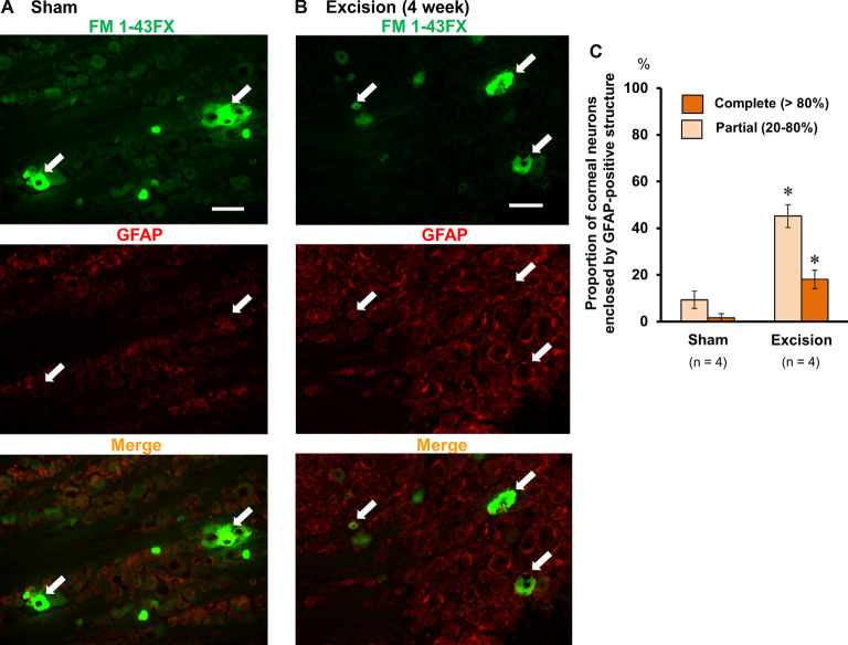
Photomicrographs of FM 1-43FX-positive corneal neurons and glial fibrillary acidic protein (GFAP)-immunoreactive cells in the anteromedial part of the trigeminal ganglion. (A,B) The trigeminal ganglion sections of the sham-operated (A) and gland excision guinea pigs (B). The corneal neurons were labeled with FM 1-43FX (green in the upper images), and the trigeminal ganglia were subsequently immunostained with anti-GFAP antibody (red in the middle images). The merged FM1-43FX/GFAP images are represented in the lower panels. The white arrows represent the FM 1-43FX-positive corneal neurons. Scale bars = 50 μm. (C) Percentage of FM 1-43FX-positive neurons completely (>80%) or partially (20–80%) enclosed by the GFAP-immunoreactive structure in the total FM 1-43FX-positive neurons. *P < 0.05 vs. the sham-operated group (Student’s t-test).

