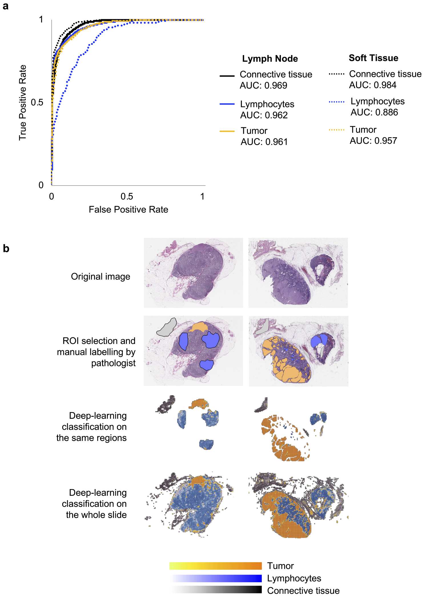
Figure 1.

Training of a Segmentation Classifier to distinguish tumor, lymphocyte, and connective tissue compartments. (a) Performance of the classifier was measured in terms of area under the curve (AUC) of the receiver operating characteristic (ROC) curve. The model performed with robust accuracy and was equally efficacious when applied to lymph node and subcutaneous tissue samples. (b) Representative images of the computational workflow. In the first row, there are two WSIs of H&E stained tissue from lymph nodes infiltrated with melanoma. In the subsequent rows, the images show manual annotation for the three regions of interest (ROI) by our pathologist coinvestigator, then training of the neural network classifier on the annotated regions, and finally, application of the classifier to the whole slide images.
