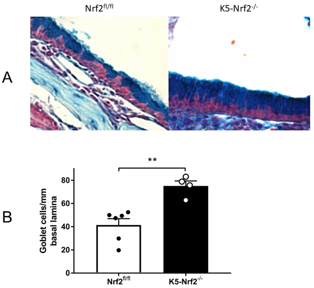
Figure 3:

Goblet cell hyperplasia in K5 Nrf2−/− sinonasal mucosa (20X). (A) Representative photomicrograph of alcian blue stained histological sections from Nrf2 flox/flox (left) or K5 Nrf2−/− mice treated with papain (right). Note increased goblet cells in the right side (B) Goblet cells were counted along the nasal septum and normalized per mm of basal lamina. Data is presented as mean±SEM. **p<0.01.
