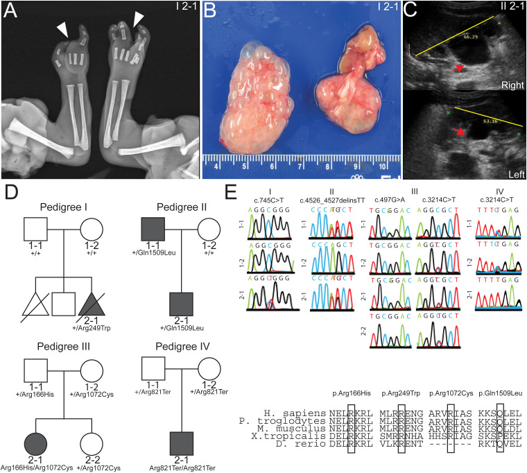
Figure 1.
Whole exome sequencing identifies discs large 5 (DLG5) variants in patients. (A) Radiograph of patient I 2–1 fetal upper extremities reveals bilateral ectrodactyly. (B) I 2–1 fetal kidneys were largely cystic and dysplastic. (C) Ultrasound of II 2–1 kidneys show hydronephrosis. Yellow line depicts span of kidney and red arrows indicate dilated renal pelvis. (D) Pedigrees depict families in which the DLG5 variants were identified. (E) Sanger sequencing confirming variant allele presence and amino conservation through phylogeny for the mutated allele of DLG5.

