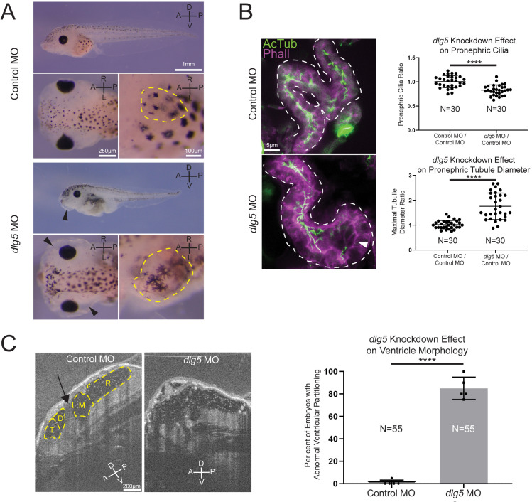
Figure 2.
Knockdown of discs large 5 (dlg5) in Xenopus embryos causes kidney and brain ventricle dysmorphology. (A) Representative images of stage 45 embryos injected with either a standard control MO or dlg5 MO show oedema (black arrow head) and kidney dysplasia (outlined in yellow) resulting from dlg5 knockdown. (B) Representative images of control MO and dlg5 MO-injected sides of stage 45 embryos along with quantitation reveal a loss of cilia and increased proximal tubule diameter in the pronephroi. Each data point is the ratio within a single embryo of either cilia detected or maximal tubule diameter in the proximal tubule between two sides either both treated with control MO (Control MO/Control MO) or one treated with dlg5 MO and the other treated with control MO (dlg5 MO/Control MO). (C) Representative images and quantitation of stage 45 embryos injected with either a standard control MO or dlg5 MO imaged via optical coherence tomography demonstrate the ventricular dysmorphology that results from dlg5 knockdown. The arrow designates the typical division between ventricles that can be found lateral to the brain midline. Embryonic axes are labelled for reference. A, anterior; D, dorsal; P, posterior; V, ventral; L, left; R, right. Ventricles are labelled according to the adjacent subdivisions of the embryonic brain. T, telencephalon; D, diencephalon; M, mesencephalon; R, rhombencephalon. Statistical tests carried out as two-tailed t-tests with ****p<0.0001 (A) or an unpaired t-test with ****p<0.0001 (C). Bars indicate mean and SD of individual values (B) or replicate means (C).

