Figure 5.
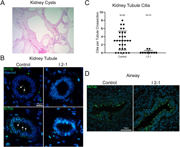
Proband tissue bearing discs large 5 (DLG5) variant c.745C>T (p.Arg249Trp) displays tissue-specific deficits in ciliation. (A) Representative H&E stain of I 2–1 kidney showing cystic morphology. (B) Control 23 weeks and I 2–1 weeks fetal kidney tissue cross-section immunofluorescence labelling of cilia Arl13b or acetylated tubulin show a loss of cilia in DLG5 variant tissue. (C) Quantitation of cilia number in each cross-sectional kidney tubule analysed. (D) Control 23 weeks and I 2–1 weeks fetal lung airway tissue cross-section immunofluorescence labelling of cilia acetylated tubulin shows intact airway ciliation in DLG5 variant tissue. Bars indicate mean and SD of individual counts.

