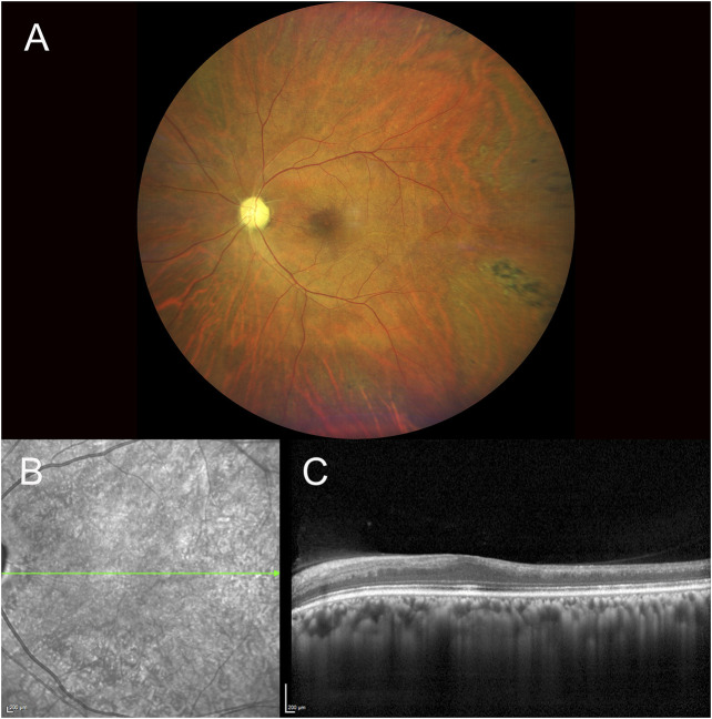
FIGURE 1.
Fundus photography of the left eye showing the presence of a pale optic disc, diffuse arterial narrowing, a mild ‘cherry-red spot’ macula and peripheral areas of retinal pigmented epithelium hyperpigmentation (A). IR and SD-OCT acquisition over the macular region of the same eye denoting atrophy of the inner retina layers with loss of foveal depression and temporal macular thinning (B,C) [IR, infrared reflectance, SD-OCT, spectral domain optical coherence tomography].

