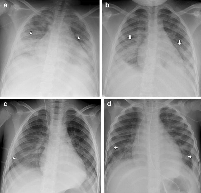
Fig. 2.
Chest radiographs of children with multisystem inflammatory syndrome in children (MIS-C). a Anteroposterior (AP) chest radiograph in a 12-year-old boy with MIS-C demonstrates bilateral consolidative and ground glass opacities (arrows) in a peripheral lower zone distribution. Categorization: typical (based on bilateral peripheral opacities). Severity score: 4. b AP chest radiograph in a 10-year-old girl demonstrates bilateral consolidative and ground glass opacities (arrows) in a peripheral lower zone distribution. Categorization: typical. Severity score: 4. c AP chest radiograph in a 4-year-old boy demonstrates a right-side pleural effusion (arrow), a right-side ground glass opacity and a left-side consolidative opacity. The opacities are in a lower zone distribution. Categorization: atypical (based on the presence of a pleural effusion). Severity score: 4. d AP chest radiograph in a 3-year-old boy demonstrates bilateral pleural effusions (arrows) and bilateral ground glass opacities in a peripheral lower zone distribution. Categorization: atypical (based on the presence of a pleural effusion). Severity score: 4

