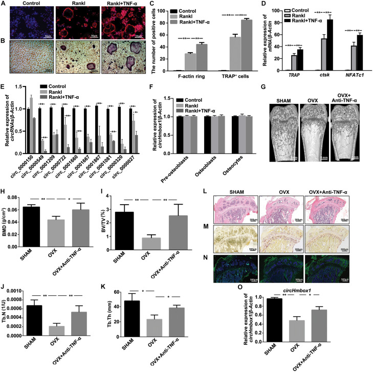
FIGURE 1.
CircHmbox1 (circ_0000549) expression is decreased in the induction of osteoclasts differentiation by TNF-α. (A) F-actin ring staining was showed in BMMs treated with or without TNF-α under RANKL and MCSF for 7 days. Scale bars are 50 μm. (B) TRAP staining was showed in BMMs treated with or without TNF-α under RANKL and MCSF for 7 days. Scale bars are 50 μm. (C) The number and area of F-actin ring and TRAP+ cells were counted. n = 4, **P < 0.01. (D) The expressions of osteoclasts formation specific genes, TRAP, ctsk and NFATc1, were analyzed by qRT-PCR in BMMs treated with or without TNF-α under RANKL and MCSF for 7 days. n = 4, **P < 0.01. (E) The expressions of circRNAs-associated osteoclastogenesis were analyzed by qRT-PCR in BMMs treated with or without TNF-α under RANKL and M-CSF for 7 days. n = 4, *P < 0.05, **P < 0.01. (F) The expressions of circHmbox1 were analyzed by qRT-PCR in pre-osteoblasts, osteoblasts and osteocytes. n = 3. (G) Representative micro-CT three-dimensional reconstructed images from SHAM mice, OVX mice and anti-TNF-α-treated OVX mice. (H–K) BMD, BV/TV, Tb.N and Tb.Th in the region of interest were measured. n = 6 mice per group. (L) HE staining was performed to histologically identify structures of the distal end of intact tibias in SHAM mice, OVX mice and anti-TNF-α-treated OVX mice. (M) TRAP staining was showed in the metaphyseal area of tibias bone sections derived from SHAM mice, OVX mice and anti-TNF-α-treated OVX mice. n = 4. (N) The immunofluorescence staining showed that anti-TNF-α alleviated OVX-inhibited osteoblastic marker osteopontin expression (green fluorescence) in the bone trabecula surface. n = 4. (O) The expression of circHmbox1 was analyzed by qRT-PCR in osteoclasts from SHAM mice, OVX mice and anti-TNF-α-treated OVX mice. n = 4, *P < 0.05, **P < 0.01.

