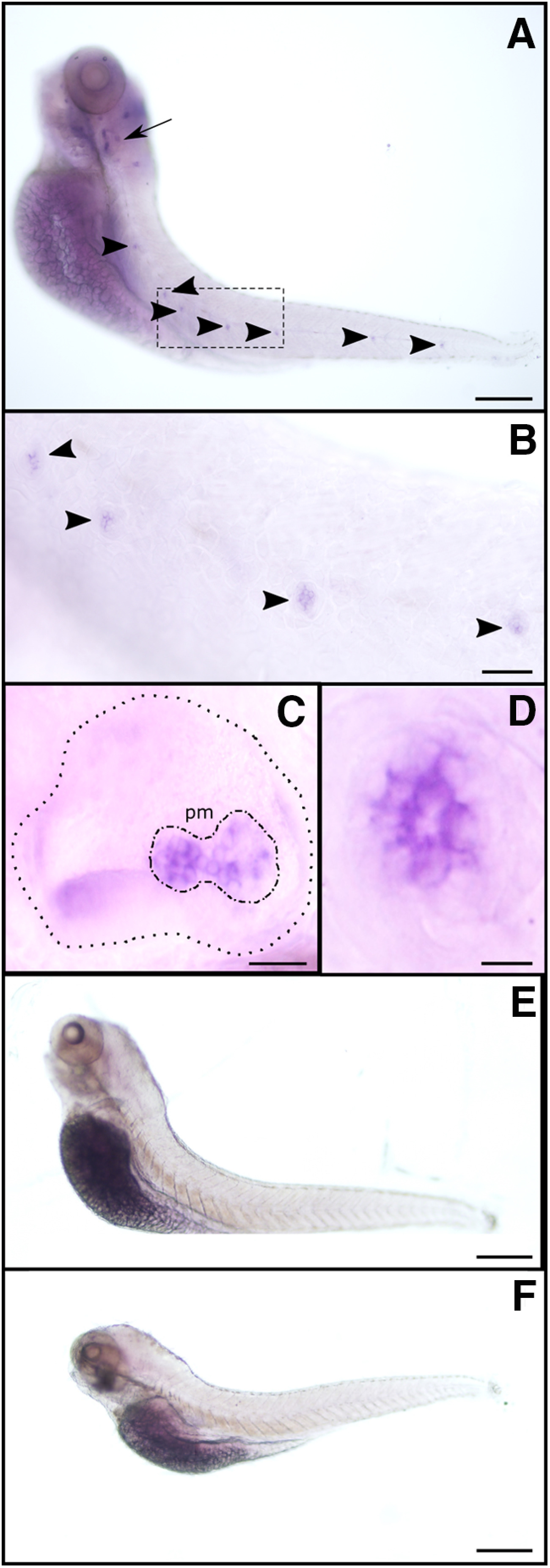
Figure 1.
ɑ9 (but not ɑ10) is expressed in zebrafish LL neuromasts and the posterior macula in the otic vesicle. A–F, Whole-mount in situ hybridization with antisense (A–D) and sense (E) ɑ9, and antisense ɑ10 (F) riboprobes. Representative lateral views, with anterior to the left and dorsal to the top, are shown. Arrow indicates the otic vesicle, and arrowheads point to selected neuromasts. B–D, Large-scale view of the otic vesicle (C) and neuromasts (B, D). C, Dotted line delimits the otic vesicle; dotted-dashed line outlines the posterior macula (pm). Scale bars: in A, E, F, 100 µm; B, 40 µm; C, 25 µm; D, 10 µm.

