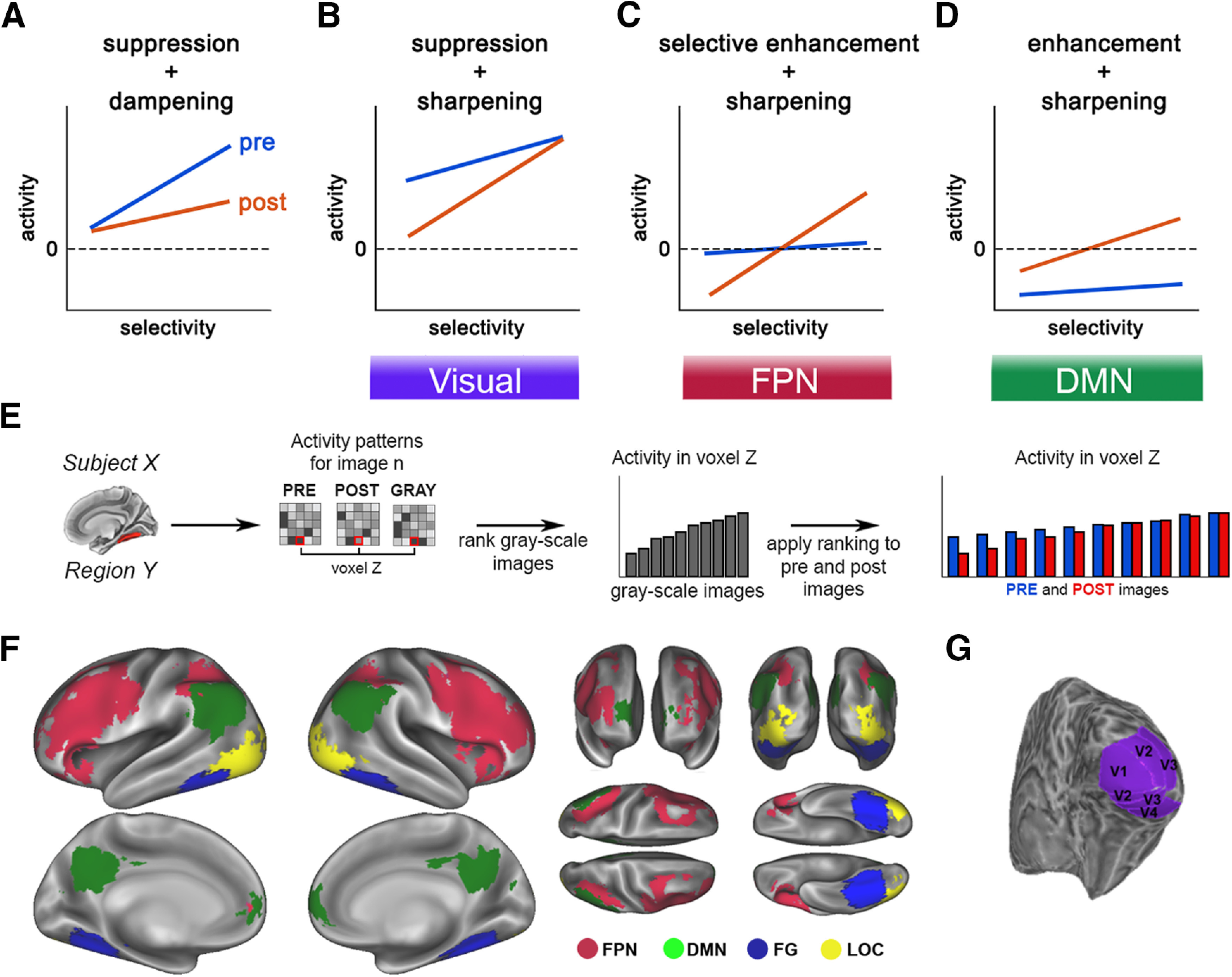
Figure 1.

A–D, Alternative explanatory mechanisms of prior-induced changes in neural activity. E, Schematic for the image preference analysis. For each voxel within an ROI, images are ranked based on the activation magnitudes in the grayscale condition. This image ranking is then applied to the same voxel's activity during pre- and post-disambiguation conditions (for details, see Image preference analysis). F, Locations of FPN and DMN ROIs (red and green), and category-selective visual areas (FG and LOC, blue and yellow). G, Location of early visual areas in an example subject.
