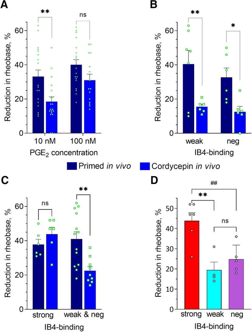
Figure 6.
Effect of in vivo treatment of fentanyl-primed rats with the reversal agent for Type I priming on PGE2-induced sensitization of small DRG neurons. Rats were primed by the systemic administration of fentanyl (30 µg/kg, s.c.) 8 d before preparing neuronal cultures (4 d before intrathecal cordycepin, followed by an additional 4 d before culture preparation). Recordings were made in small-diameter DRG neurons from fentanyl-primed animals either treated (reversal group) or not treated (primed group) with cordycepin (depicted in A–C by the blue and dark blue bars, correspondingly) after 24 h in culture. In A–D, bars show pooled magnitudes of decrease in rheobase, relative to baseline, after PGE2 application (10 and 100 nm), measured and analyzed in the same way as described in Figure 5 and Materials and Methods. Symbols show individual values. In A–C, values for primed group were repeated from Figure 5A–C for the purpose of comparison. A, Reduction of rheobase in response to 10 and 100 nm PGE2, analyzed regardless of IB4-binding status. In neurons from primed rats the effect of 10 nm but not 100 nm PGE2 was significantly greater than in the reversal group [two-way ANOVA; effect of condition: F(1,68) = 12.5, p = 0.0007; Holm–Sidak's post hoc: t(68) = 3.2, **adjusted p = 0.004 for 10 nm; t(68) = 1.8, adjusted p = 0.07, not significant (ns), for 100 nm]. Number of cells in primed group: n = 19 for both 10 and 100 nm; in reversal group: n = 19 for 10 nm and n = 15 for 100 nm. B, Reduction of rheobase in response to 10 nm PGE2 in weakly IB4+ (“weak”) and IB4– classes (“neg”) neurons. Attenuation of PGE2-induced sensitization after in vivo cordycepin was statistically significant in both neuronal populations (two-way ANOVA: effect of condition F(1,21) = 16.3, p = 0.0006; Holm–Sidak's post hoc: t(21) = 3.2, **adjusted p = 0.008 for weakly IB4+; t(21) = 2.5, *adjusted p = 0.02 for IB4–). Number of cells (weak/neg) in reversal group: 6/6, in primed group: 7/6. C, Reduction of rheobase in response to 100 nm PGE2 in strongly IB4+ (“strong”) and merged weakly IB4+ and IB4– (“weak and neg”) neurons from primed and reversal groups. Two-way ANOVA revealed statistically significant interaction (F(1,30) = 8.5, p = 0.007), indicating differential effects on the three different neuronal classes. Indeed, statistically significant attenuation in reversal compared with primed group occurred in weakly IB4+ and IB4– but not in strongly IB4+ neurons (Holm–Sidak's post hoc: t(30) = 0.9, adjusted p = 0.38 for strong, not significant (ns); t(30) = 3.7, **adjusted p = 0.002 for weak and neg). Number of cells (strong/weak and neg): 6/13 in primed group, 6/9 in reversal group. D, Reduction of rheobase in response to 100 nm PGE2 in strongly IB4+ (strong), weakly IB4+ (weak), and IB4– (neg) neurons from reversal group. Values in strongly IB4+ class were significantly greater than in weakly IB4+ and IB4– neurons [one-way ANOVA: F(2,12) = 11.8, ** p = 0.002; Tukey's post hoc: q(12) = 6.3, adjusted p = 0.002 for strongly IB4+ vs weakly IB4+; q(12) = 5.2, ##adjusted p = 0.008 for strongly IB4+ vs IB4–; q(12) = 1.3, adjusted p = 0.63, not significant (ns), for weakly IB4+ vs IB4–]. Number of cells: 6 strong, 4 weak, 5 neg.

