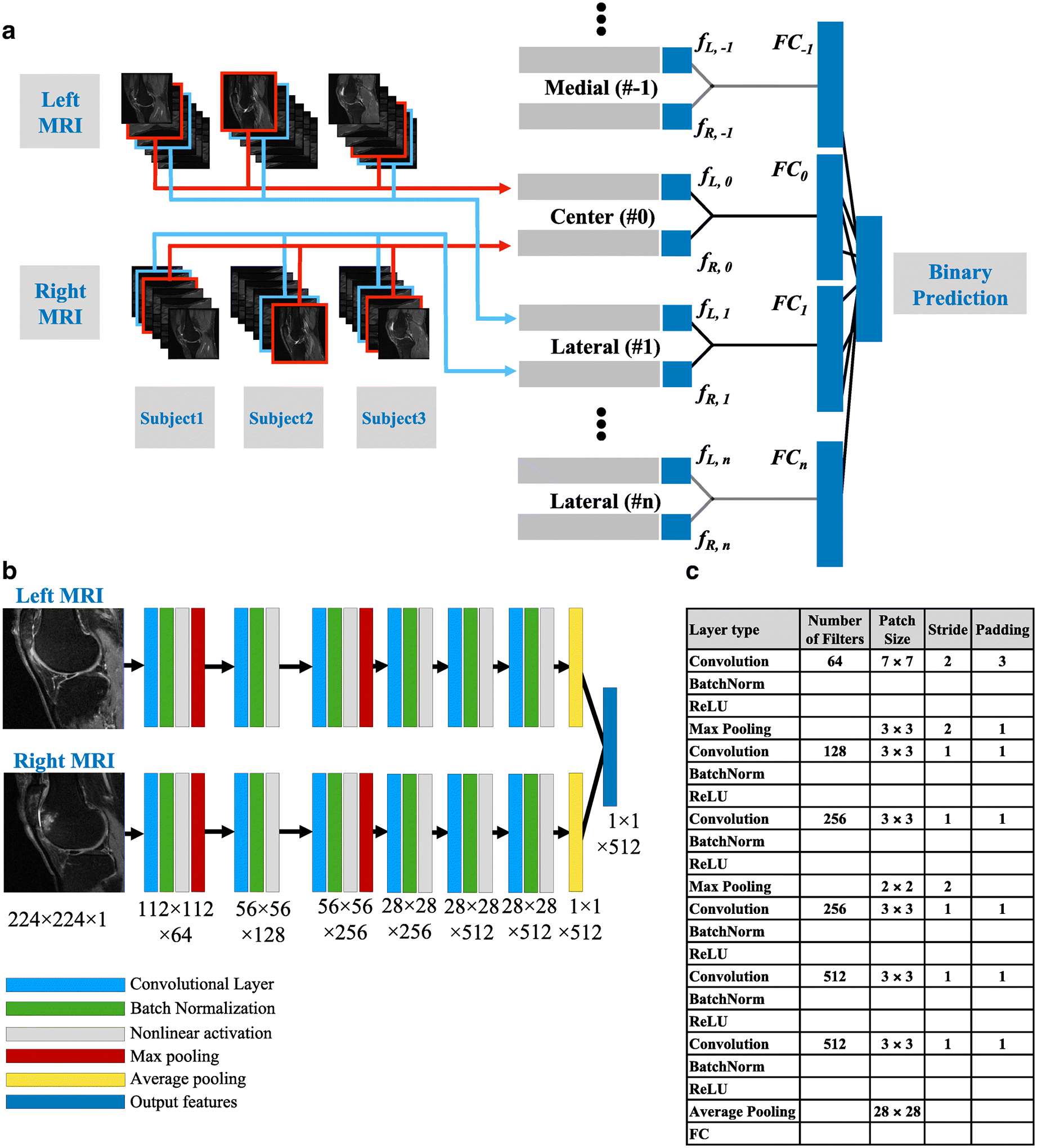
Figure 2: Convolutional Siamese network architecture.

(A) After image pre-processing, the indexed MRI slices were fed into the corresponding Siamese network model. The outputs from all the 2D models were concatenated for the binary prediction task. (B) The 2D MR slices for the left and right knee were fed into the network simultaneously. Each neural network that is part of the Siamese architecture comprised of 6 convolutional layers. Black arrows represent connection between the images and the first layer, connections between the layers and the connection between the layer and the output. For layers 1 and 3, each convolutional operation was followed by batch normalization, nonlinear activation and max pooling, whereas for layers 2, 4, 5 and 6, each convolutional operation was followed by batch normalization and nonlinear activation. Only the first convolutional layer and the two max pooling layers had a stride of 2, whereas the other layers had a stride of 1. Consequently, the final convolutional layer had a high in-plane resolution with a dimension of 512×28×28. (C). Table presenting the details of the different layers within the neural network.
