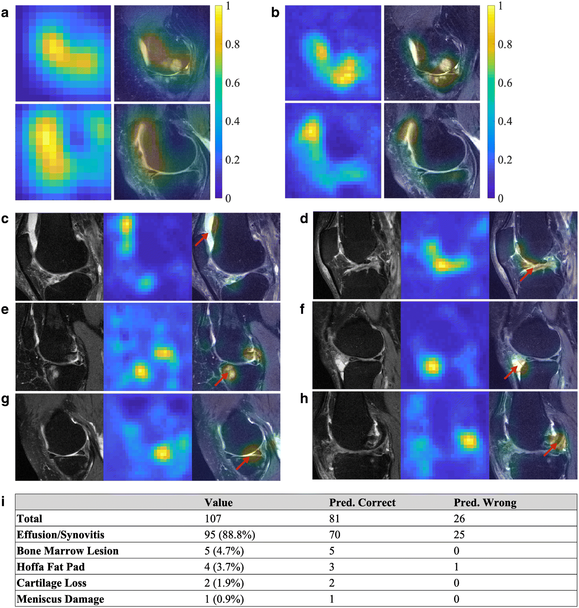
Figure 3: CAMs on selected subjects within the test data.

(A) Examples of CAMs generated from the fine-tuned VGGNet model, resulting in CAMs with an in-plane resolution of 14×14 pixels. (B) Examples of CAMs generated from the present model from the same MRI images with an in-plane resolution of 28×28 pixels. Both the heat maps and the overlap of the MR image with the heat map are shown. In some cases of the test data (C, D), effusion/synovitis was identified as the lesion present within the hot spots. Also, in few other cases, (E) bone marrow lesions, (F) Hoffa fat pad abnormality, (G) cartilage loss, and (H) meniscus damage, were identified as the lesions present within the hot spots. The red arrows indicate the locations of the identified structural regions. (I) Radiologist’s assessment on the test cases (n=107). For each case, the MR scan and the model-derived heat map of the knee with confirmed pain were reviewed by the radiologist, who then identified the presence of any lesions within the regions highlighted by the heat map.
