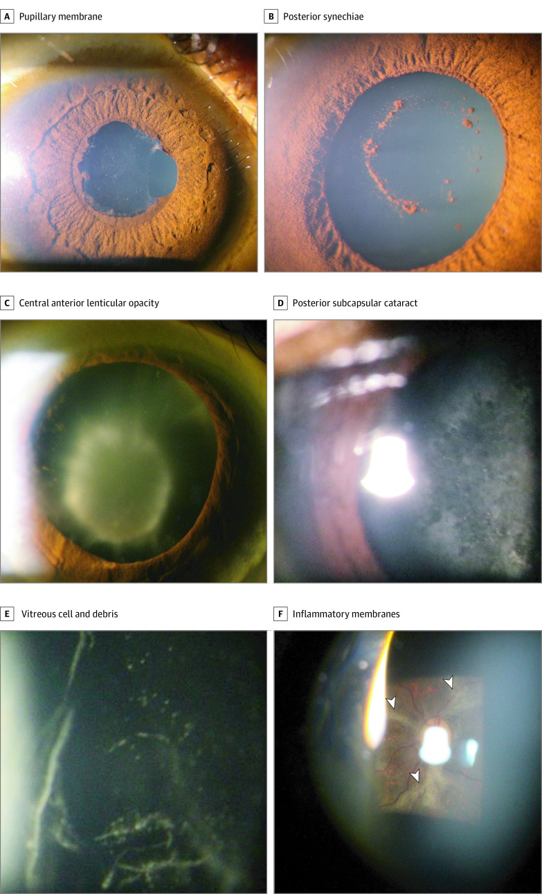
Figure 2. Slitlamp Biomicroscopic Examination of Eyes Affected by Ebola Virus Disease.
A, Posterior synechiae and pupillary membrane appeared in an eye with anterior uveitis. B, Pigment on the anterior lens capsule remained after lysis of synechiae with cycloplegic drops. C and D, Inflammatory cataracts in eyes with a history of uveitis included central, anterior lenticular opacity (C), and posterior subcapsular cataract (D). E, Vitreous cells and debris appeared in an eye with intermediate uveitis. F, As imaged through a 90-D lens, gliotic processes (arrowheads) are seen extending from the optic nerve in the eye of a survivor of Ebola virus disease.

