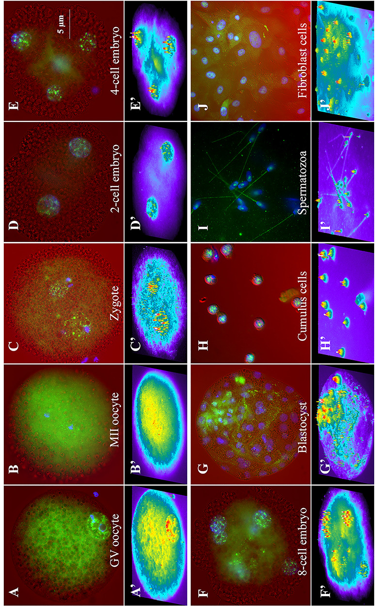
Figure 4.

Representative immunofluorescence images of NEDL2 (green) in porcine ova, zygote, and embryos generated by IVF. NEDL2 labeled in green, and DNA counterstained with DAPI in blue were superimposed over the corresponding light-microscopic images in red acquired with differential interference-contrast (DIC) optics (on top, A–J). The intensity of NEDL2 expression was further analyzed by MetaMorph software (v7.1) and presented as intensity profiles in the bottom panels (A” to J”) using heat map pseudocoloring. The following oocyte, embryos, and cell types are shown: (A) GV-stage oocyte, (B) metaphase II oocyte, (C) 30 h post-IVF zygote, (D) 2-cell stage, (E) 4-cell stage, (F) 8-cell stage, (G) day 7 blastocyst stage embryo, (H) surrounding GV-stage-oocyte cumulus cells, (I) d35 fetal fibroblast cells, and (J) ejaculated spermatozoa. Typical, representative patterns of NEDL2 labeling are shown (color merged A’–J’). NEDL2 showed both a diffused cytoplasmic localization and accumulation in distinct foci in the GVs, pronuclei, or nuclei at all stages of oocytes and embryos and in fibroblast and cumulus cells. Ab154888 antibody was used in ICC analysis.
