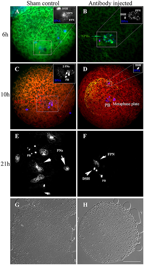
Figure 5.

Representative images of oocytes at 6, 10, and 21 h after IVF in normal serum (sham) and anti-NEDL2 antibody (ab154888) injected groups. DNA was counterstained with DAPI (blue). (A, B) Immunofluorescence of NEDL2 (green) in the sham and anti-NEDL2 antibody-injected oocytes at 6 h post-IVF. It also illustrates that the anti-NEDL2 antibody injection accelerated the formation of nucleolus precursor bodies. Spermatozoa were labeled with MitoTracker in red. (C, D) Immunofluorescence of injected anti-NEDL2 antibody (green) and oocyte endogenous NEDL2 (red) at 10 h post-IVF. The injected antibody was detected and labeled in green and red, and shown in orange in (D) after merging the two colors. (E, F) Comparison of pronuclei formed at 21 h after IVF. Multiple pronuclei formed in the sham control, while only female pronucleus formed at this time, and sperm DNA was still not decondensed. (G, H) The corresponding light-microscopic images acquired with differential interference-contrast (DIC) of sham and antibody injected zygotes. Note: the sperm DNA did not decondense in the antibody injected group at 6, 10, and 21 h after IVF. DSH: decondensing sperm heard; NPB: nucleolus precursor body; PB: polar body; PN(s): pronucleus (pronuclei); FPN: female pronucleus; MPN: male pronucleus. Bar = 25 μm.
