Figure 2.

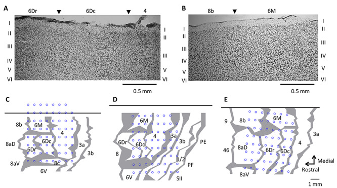
(A–B) Parasagittal sections illustrating cytoarchitectural characteristics of premotor (6Dr and 6Dc), supplementary motor (6M) and primary motor (4) cortex. Panels A and B respectively represent sections from MK2 and MK3. C-E. Reconstruction of array location over the frontal motor cortex by histological assessment for MK1 (C), MK2 (D), and MK3 (E). The top black horizontal line represents the midline. Gray lines indicate architectonic borders, reconstructed from sagittal sections. The thickness of the lines represents zones of uncertainty. Blue circles refer to the location of each of 63 recording electrodes.
